ELISA methodologies are a gold standard method for regulatory authorities for monitoring the removal of HCPs during the downstream process.
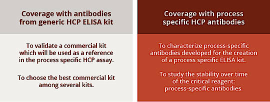
The authorities also request to perform a COVERAGE analysis of the anti-HCP antibodies contained in the HCP ELISA kit used for the assay.
This analysis, called COVERAGE, is an estimate of the percentage of HCP proteins detected (or covered) by the antibodies of the ELISA kit (process-specific anti-HCP antibody or generic anti-HCP antibody kit. Several techniques are available to set up this COVERAGE analysis and it is recommended to use 2D techniques.
Custom COVERAGE analysis by AGRO-BIO
HCP coverage evaluation : When should you do it?
Which Coverage analysis are performed in-house ?
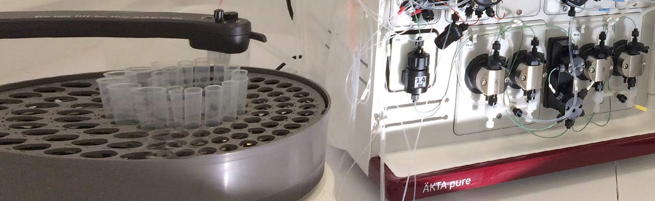
In-house, we can perform two different methods for HCP coverage evaluation: Coverage by 2D-DIBE, and Coverage by Protein Selective Purification. Both are based on a two-dimensional (2D) electrophoresis approach allowing to have on a gel an exhaustive mapping/snapshot of HCP in the form of spots (migration of proteins according to their isoelectric point (pI) and their size). These protein spots are then revealed by staining or by exploiting fluorophore labeling (Cy2, Cy3, ...). The choice of the coverage assessment method for a project is determined by our scientific team according to the context, the products availability and the objectives. For example, the measurement of coverage by HCP selective purification consumes anti-HCP antibodies, which is a limiting factor for some projects.
How is calculated Coverage percentage (%) ?
To estimate the coverage percentage we determine the number of spots present on the analyzed gels or membrane from two channels. In case of 2D-DIBE, it means first the signal from staining or labeling of total HCP then the signal from secondary Ab revealing Commercial or Process specific Ab. In case of PSP, it means first the signal from staining or labeling of total HCP then the signal from staining or labeling of Selectively purified HCP.The number of spots from both channels is assessed. For this we use the software MelanieTM9 of Cytiva®, which is a comprehensive solution for visualization, matching, detection, quantitation, and analysis of 2-D gel and membraneimages. It was specially designed for coverage analysis, to monitor HCP impurities in biopharmaceutical products.
Thanks to its 3D view MelanieTM9 minimizes false positives spots, it also monitor the robustness of the assay with Coverage Range, and evaluate the performance of anti-HCP Antibodies with automated quadrant coverage detection. Authorities require maximum coverage and sensitivity against native HCP for the building of a suitable ELISA kit, with equal distribution over the 4 quadrants of the gel.